Labelled Pictures Of Human Skin : Skin diagram labeled - Sensory receptors in the human skin.
Labelled Pictures Of Human Skin : Skin diagram labeled - Sensory receptors in the human skin.. Related posts of labelled diagram of a human skin anatomy human body organs female. Click on the tags below to find other quizzes on the same subject. A keratinocyte is a cell that manufactures and stores the protein keratin. Eczema is the most common form of skin disease in humans. Discover (and save!) your own pins on pinterest
Take the skin diseases pictures quiz and learn to identify common conditions that plague human skin. Skin also helps maintain a constant body temperature. Also explore over 33 similar quizzes in this category. Undoubtedly, the skin is the largest organ in the human body; Eczema is the most common form of skin disease in humans.
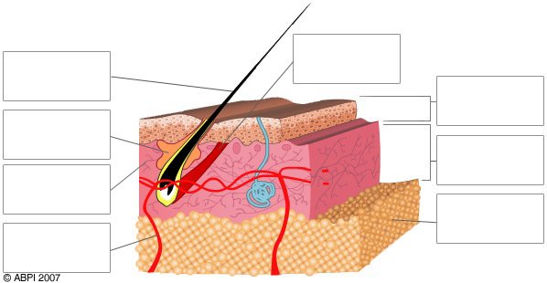
Skin - Merocrine Sweat Glands - Histology from i.pinimg.com The human skin is the outer covering of the body and is the largest organ of the integumentary system.the skin has up to seven layers of ectodermal tissue and guards the underlying muscles, bones, ligaments and internal organs. Sensory receptors in the human skin. Eczema is the most common form of skin disease in humans. In this quiz, we are going to focus on the skin structure of the human body. The epidermis, an outermost layer that contains the primary protective structure, the stratum corneum; The dermis, a fibrous layer that supports and strengthens the epidermis; Though nearly all human skin is covered with hair follicles, it can appear. The human skin is composed of three layers of tissue:
Oral health explore images of dental and oral health diseases as well as cosmetic dentistry before and after pictures.
Also explore over 33 similar quizzes in this category. It is a tough protective layer that contains melanin (which protects against the rays of the sun and gives the skin its color). The epidermis, an outermost layer that contains the primary protective structure, the stratum corneum; This is an online quiz called label the skin. Sensory receptors in the human skin. When ultraviolet light waves touch melanocytes, they begin to increase the production of melanin. Literally covering you from head to toe. It's easy to take your skin for granted, but when you consider how it protects your body from harm, it is something we should appreciate more. This skin diagram lists all the important parts of human skin, including the dermis, epidermis, hypodermis, sweat pore, hair shaft, pigment layer, nerve fiber, dermal. This article will describe the anatomy and histology of the skin. Undoubtedly, the skin is the largest organ in the human body; The dermis, a fibrous layer that supports and strengthens the epidermis; Posted in bones, diagrams | tagged body skeleton, human skeletal anatomy, human skeleton, human skeleton anatomy, skeletal, skeletal anatomy, skeletal images, skeletal system, skeleton picture of body organs location 2.
This article will describe the anatomy and histology of the skin. This diagram depicts labeled human skeleton diagram with parts and labels. The cells in all of the layers except the stratum basale are called keratinocytes. Is a health blogger focusing on health, beauty, lifestyle and fitness topics. Printouts label human anatomy diagrams.
Labeled Picture Of The Nervous System Peripheral Nervous ... from i.pinimg.com Printouts label shapes, food, the body, and other things in italian. The human skin is the outer covering of the body and is the largest organ of the integumentary system.the skin has up to seven layers of ectodermal tissue and guards the underlying muscles, bones, ligaments and internal organs. This is an online quiz called label the skin. To do so, we need to determine the number of skin regions in the image. Select from premium human skull of the highest quality. Picture of human body with organs labeled, download this wallpaper for free in hd resolution.picture of human body with organs labeled was posted in june 11, 2017 at 5:58 am. Human skin is only about 0.07 inches (2 mm) thick. Skin also helps maintain a constant body temperature.
Human skin is only about 0.07 inches (2 mm) thick.
Printouts label shapes, food, the body, and other things in italian. Desquamation (sloughing of cells) from the epidermis, thick skin, human, 100x at 35mm. The human skin is the outer covering of the body and is the largest organ of the integumentary system.the skin has up to seven layers of ectodermal tissue and guards the underlying muscles, bones, ligaments and internal organs. Take the skin diseases pictures quiz and learn to identify common conditions that plague human skin. He has been with healthiack.com since 2012 and has written and reviewed well over 500 coherent articles. Literally covering you from head to toe. Click on the tags below to find other quizzes on the same subject. It does not contain blood vessels. Picture of human body with organs labeled, download this wallpaper for free in hd resolution.picture of human body with organs labeled was posted in june 11, 2017 at 5:58 am. When ultraviolet light waves touch melanocytes, they begin to increase the production of melanin. The skin becomes dark color. Its color boundary is represented by pixels with value 1 for binary images. This is an online quiz called label the skin.
A skin region is defined as a closed region in the image, which can have 0, 1 or more holes inside it. The dermis, a fibrous layer that supports and strengthens the epidermis; Skin diseases like eczema, if not detected and controlled early, may lead to severe health and financial consequences for patients. There is a printable worksheet available for download here so you can take the quiz with pen and paper. Beneath the two layers is a layer of subcutaneous fat, which also protects your body and helps you adjust to outside temperatures.
skin blank labelled : Biological Science Picture Directory ... from pulpbits.net The epidermis, an outermost layer that contains the primary protective structure, the stratum corneum; A skin region is defined as a closed region in the image, which can have 0, 1 or more holes inside it. A keratinocyte is a cell that manufactures and stores the protein keratin. Skin is made up of two layers that cover a third fatty layer. Detailed texture of human skin. It's easy to take your skin for granted, but when you consider how it protects your body from harm, it is something we should appreciate more. In addition to human anatomy and physiology, this collection of images illustrates many of the major diseases and conditions of the body. Don't forget to share this picture with others via.
Literally covering you from head to toe.
Human skin is only about 0.07 inches (2 mm) thick. Description from picture of human body with organs labeled pictures wallpaper : Printouts label musical notes, rests, notation, keyboards, and scales. It does not contain blood vessels. It's easy to take your skin for granted, but when you consider how it protects your body from harm, it is something we should appreciate more. Detailed texture of human skin. Desquamation (sloughing of cells) from the epidermis, thick skin, human, 100x at 35mm. It is a tough protective layer that contains melanin (which protects against the rays of the sun and gives the skin its color). Oral health explore images of dental and oral health diseases as well as cosmetic dentistry before and after pictures. Related posts of labelled diagram of a human skin anatomy human body organs female. This hd wallpaper picture of human body with organs labeled has viewed by 1011 users. Discover (and save!) your own pins on pinterest A keratinocyte is a cell that manufactures and stores the protein keratin.
0 Response to "Labelled Pictures Of Human Skin : Skin diagram labeled - Sensory receptors in the human skin."


0 Response to "Labelled Pictures Of Human Skin : Skin diagram labeled - Sensory receptors in the human skin."
Post a Comment